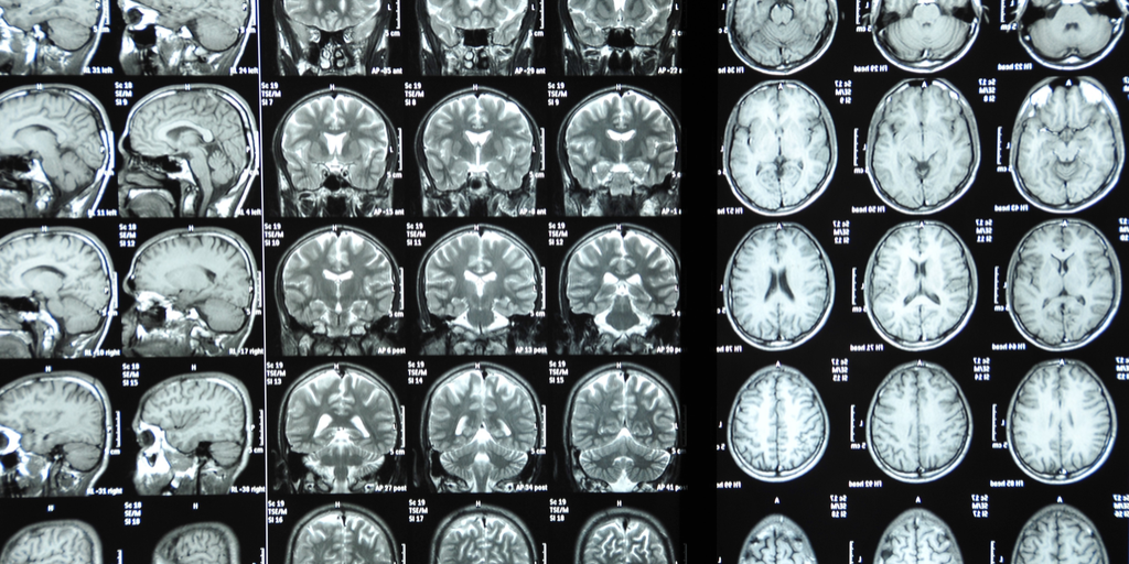
I finished my second MRI scan this year earlier this afternoon. So far lifetime – I have had 5 and will have 6 by the end of next month. I am sure that many people reading this have had the experience and I would not rate it as pleasant at all. Just the obsessive checklist that must be completed prior to the scan is enough to raise the anxiety level. Is my body free of implanted or tattooed or accidentally placed metals? When they were scanning my pancreas – my first thought was: “What about that laparoscopic cholecystectomy I had done in 2019?” I had read the operative report and it described two permanent clips being placed on the cystic duct prior to dividing it and removing the gallbladder. I contacted the surgeon about that and he was certain the clips were not ferromagnetic.
One of the last questions is: “Are you claustrophobic?” And if you are is your primary care doctor prescribing
a sedative and if you take that sedative is there somebody here who can drive
you home?” I would be hard pressed to think of many people who would not be
claustrophobic in an MRI tube. After all
you are in a tight space with very loud noises for a prolonged period. As the
radio frequency waves are generated the tube heats up. It is better than hurtling through space in a
larger tube during air travel – and I smiled to myself as I thought of the
comparison.
In all cases I have been given a headset and asked about
musical preferences and volume. So far,
the headsets don’t block the sound of the machine and obviously are not noise
cancellation devices. It did lead me to think about designing headsets without
ferromagnetic materials and what that might involve. The only designs I have
seen so far use air conduction through tubes rather than electrical
connections.
Music selection is as much of a problem as the low-fi
headset. I forgot to ask if I could use
my own playlist – but it was safely locked up far away from the 1.5T magnet. I
tried to be more specific this time: “Have you got any Canned Heat?” My most recent play list starts with 3 Canned
Heat songs from 1967 – but looking at the tech I estimated he was born in
1980 and the other in the 1990s. When they
rolled me out of the tube one had been replaced by a woman with brightly colored
hair who may have been born in the 21st century. I changed my answer to “Classic rock.” That can be a disappointing genre because too
much of it is bubble gum music. Over the
30 minutes in the tube, the heaviest it got was AC/DC Highway to Hell and
James Gang Funk 49. I did
enjoy Steve Perry (Journey) and
was tempted to sing along like I do in the car.
But I am sure that would have not been a good idea and may have resulted
in additional imaging time and I can no longer hit the high notes.
My mind wandered to fMRI research. How in the world can
research subjects be expected to produce real world results from inside the
catastrophic MRI world? I decided not to include my real catastrophic thinking in this post because it is idiosyncratic and I don't want to affect anyone else's decision to get an MRI scan. And today I just had one or two brief thoughts. I spend most of
the time in the tube actively distracting myself and doing sigh breathing
exercises to control my heart rate. Today
I opened my eyes in the tube – briefly for the first time. All I could see was an expanse of whiteness
in front of my face with a row of fasteners bisecting the field. I was pleasantly surprised to find it was
about 6 inches away – farther than I had imagined it.
While I was thinking about research, I also thought about all of the MRI scans I had ordered on my patients. Going through the procedure yourself leads to questions about the how it is presented to patients for informed consent. I was careful to describe the issues with confined space and noise as well as the advantage of no radiation and better resolution. Being hospital based, I had the advantage of an anesthesia team being available to sedate and monitor patients who were unable to tolerate it. As I was showing one person their results by holding the film against a window in their room they fainted and I was able to catch them on the way down. The realistic appearance of the brain in that scan led to that reaction.
Forty years ago, I was an intern in this hospital. My very
first rotation was Internal Medicine. Back
in those days it was a county hospital.
Today it is a massive flagship hospital of one of the largest health
care organizations in Minnesota. That included a building program to the tune
of hundreds of millions of dollars. The original hospital remains at the
center, but it is obscured by new wings and buildings. The parking lot I parked
in did not exist at the time. There was
a lot out front that you accessed with a magnetic card. One night I was working late and a guy
approached me for money as I entered that lot. I handed him $20 to avoid what I thought might be
coming. It was a tough neighborhood.
Once you enter the building – you can step back in time
where old meets new. One of those places
is medical imaging. During internship and in the 22 years I worked there radiology
(as we used to call it) was one of my favorite haunts. I knew the radiologists
and knew I could ask them questions about films. Surprisingly many of them had questions about
psychiatry. Before the electronic health record, I would make a drawing of the
positive findings from CT and MRI scans and redraw it in the patient’s chart. As radiology became digitized it was easier to
cut and past images. I could still
discuss images with the neuroradiologist.
I missed all of that when I left that practice.
The MRI tech comes over the headphones: “OK we are going to come in and inject the
contrast.” They know I had a mild
reaction to CT scan contrast but the MRI contrast is gadolinium based and I
have had it before. The last time they
checked my pre and post creatinine levels, but at this facility that is
replaced by questions about renal insufficiency and dialysis. That seems like a high bar. He checks in again in 5 minutes to make sure
that I am not having a reaction.
Another sequence of radiofrequency waves starts and there is
a pulsating beat that reminds me of a rock and roll song. I try to recall the song just based on the
beat. I check my muscle tension and
realize my shoulders are rolled forward – so I force myself to relax, move my
neck, and do some patterned breathing. I would really like to hear some Nirvana
at this point.
The tech is on the headphones again. “OK you are doing great – 5 minutes left.” That reminded me of a previous scan when I
got a similar message and remembered all of the songs that played afterwards. This time ZZ Top LaGrange comes on. It
is a 4 minute song. At the end – they roll
me out of the tube and tell me I have done a great job.
Last night I was wondering whether I was getting
progressively more anxious about MRI scans or whether this was a form of
exposure therapy. I was surprisingly calm during this one and more confident
that I will live to MRI another day.
George Dawson, MD, DFAPA
Supplementary: I have received some early feedback on why I am getting these MRI scans. First and foremost - I am interested in addressing serious problems and preventing disability. I personally know many people who were disabled as the result of spinal injuries that occurred from seemingly trivial events like turning to see someone walking through the door or turning over in bed. I am also aware of age related injuries that occur in active people. Falling off your bike at age 70 is not the same as falling off your bike at age 30 or 40. All of these scenarios suggest to me that numerous age-related changes in the spine in the absence of any course of effective strengthening can lead to catastrophic problems.
In addition to the symptoms, I would like to get an opinion of whether it is safe to do aspects of my exercise routine. I would really like to get back out on the ice speedskating - but I am not going to if it means I will get progressively disabled from spinal problems. I saw Lindsey Vonn speak to this recently. I am certainly not comparing myself directly to one of the greatest skiers of all time - but I could relate to why she finally decided to quit.
“My body is broken beyond repair and it isn’t letting me have the final season I dreamed of,” Vonn said. “My body is screaming at me to STOP and it’s time for me to listen.”
A lot of aging athletes like myself have the thought that as long as we exercise and stay very active - we will be able to continue in sports as long as we want. In retrospect, I think I have shown that you can certainly push it much farther than expected and much farther than average - but like the best there is a breaking point.
Without yet knowing the result of this scan - the possibilities are significant. In the ideal world, it will show age related changes and that would just indicate continuing the physical therapy that I have been doing for 20 years. If a potential surgical problem shows up that is more complicated. I do know skaters who have had back surgery and most back surgery has highly variable results. I have also observed and assisted on many back surgeries in medical school where neurosurgery was my preferred surgical rotation - but I assume surgical technique has improved greatly since then. Would I get back surgery if there was a high likelihood of symptomatic relief and I could return to skating? Would I get back surgery without that guarantee? All of that is up in the air at this point.
Image Credit: Image credit and Creative Commons licensing can be obtained by clicking directly on the image at the top of this post.